Affichages
(→Dynamic and Static mode in medical thermal image) |
(→Dynamic and Static mode in medical thermal image) |
||
| Ligne 85 : | Ligne 85 : | ||
An improvement of the images can still be done to use a plate at a body level temperature in order to decrease contrast and physical crisp during thermal shoot. | An improvement of the images can still be done to use a plate at a body level temperature in order to decrease contrast and physical crisp during thermal shoot. | ||
| + | |||
| + | French version: [[Statique versus dynamique en thermographie médicale]] | ||
[[Category:Library]] | [[Category:Library]] | ||
Version du 17 mars 2019 à 13:32
Sommaire |
Dynamic and Static mode in medical thermal image
In thermography, we have the choice to observe the moment, in static, the evolution or cause ourselves the chance, that's the dynamic mode.
Dynamic mode request a broad experience in order to master the consequences of the stimulation and their limits. It requests too to catch the desired instant because dynamic is by definition fugace...
A lot of things can be observe, the change voluntary provoqued or the fall back to normality, evolution or new equilibria's point.
Observation of the change itself
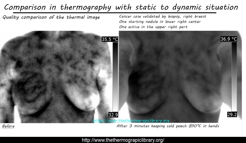

At left, original situation, static mode, inverted grey palette.
At right, dynamic observation after subject kept in her hands a gel cool pack at a temperature of around 10°C during 3 minutes.
Quality of image is completely changing and patterns are now well sharp. Of course first image allows already to analyze the situation but second is clearly easier to interprete and this finally remove minor points from the observation.
In some case, this is the only way to reveal an infection or illness who's quite discrete without this kind of protocol.
Post-observation, the restore to the "normality"

Material is here the same, a classical coolpack containing gel with a temperature of around 10°C, kept in hand during 30 seconds.
One major point is coolpack must absolutely be dry!! That's the Reason why we don't use simply water, to avoid the effect of water on skin who will polluting the observation, covering it in fact.
This protocol can replace an heavy examination with echography combinated with Larsen. the aim is studying the global vascularization by checking restoration time and his level on the hand after a cooling.
Source of the protocol: Dual-mode Imaging of CutaneousTissue Oxygenation and Vascular Function
Protocol case with static cold and hot stimulation
We will here use gel pouches to avoid to wet the skin with a temperature of around 5°C during 2 minutes, a little pause and next using a massaging device (image is high resolution, each thermal scale is readable, click on it) .

Which conclusion could we have after this experiment?
1 = Static situation, start and 2 = dynamic, final situation. This is performed in an uncontrolled area, by choice.

Results are impressive, we see that despite the thermal contrast decrease, image is far more readable. Worst, that pain area was not readable in static mode but perfectly displayed after dynamic stimulation.
And, finally, here is the precision of the result we can obtain with a focusing on the middle left of the last articulation as the source of the problem:

We can see that middle finger have a light problem but that index's last articulation demonstration a problem with light inflammatory at left middle of it. The thermography can allow here to identify location and improve diagnose with additional information.
Additional remark
An improvement of the images can still be done to use a plate at a body level temperature in order to decrease contrast and physical crisp during thermal shoot.
French version: Statique versus dynamique en thermographie médicale