Affichages
| Ligne 9 : | Ligne 9 : | ||
| − | One of the classic calf ailments are | + | One of the classic calf ailments are varicosity (small intradermal dilations under 3 mm) but may be the beginnings of much more troublesome varicose up even painful phlebitis, medical world have not the same approach regarding the location as for anglo-saxon world and America varicosity is mitigated into varicose veins but in continental Europe they can be separated as varicosity is considered as mild. |
Diagnosis is currently done primarily by Doppler ultrasound (ultrasonography) but thermography allows its visualization even though it is mainly useful for darker skin where direct visualization is difficult or delayed by the less transparency of the skin. | Diagnosis is currently done primarily by Doppler ultrasound (ultrasonography) but thermography allows its visualization even though it is mainly useful for darker skin where direct visualization is difficult or delayed by the less transparency of the skin. | ||
Version du 5 août 2015 à 14:08
Varicose veins in thermography
Calf is the back portion of the lower leg into the human body. The muscles within the calf correspond to the posterior compartment of the leg. The two largest muscles within this compartment are known together as the calf muscle and attach to the heel via the Achilles tendon. Several other, smaller muscles attach to the knee, the ankle, and via long tendons to the toes.[1]
Can be very muscular, calf plays an essential role in walking because the contraction of his muscles will be used to pump the blood stream.
Once the calf was seen in some cultures as a sign of strength to the point that some were adding padding to be better preceived; ironically, it is actually correlated that having thin calves could meanto health problems in old age on malnutrition and carotid plaques (part of arteriosclerosis) [2].
One of the classic calf ailments are varicosity (small intradermal dilations under 3 mm) but may be the beginnings of much more troublesome varicose up even painful phlebitis, medical world have not the same approach regarding the location as for anglo-saxon world and America varicosity is mitigated into varicose veins but in continental Europe they can be separated as varicosity is considered as mild.
Diagnosis is currently done primarily by Doppler ultrasound (ultrasonography) but thermography allows its visualization even though it is mainly useful for darker skin where direct visualization is difficult or delayed by the less transparency of the skin.
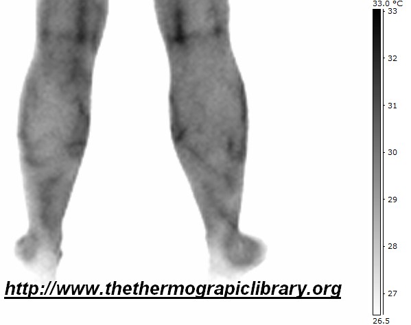
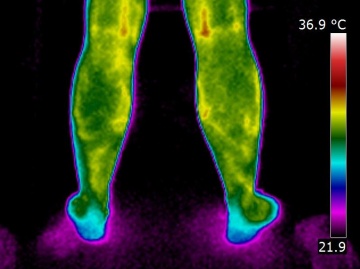
Calves suffering of simple varicosities but can be a warning signal for a later varicose veins evolution.
Here is now a case of varicose veins compared to the previous spider veins case and on the right a flebitis state:
Varicose veins upper ------------------------------------------------------------------------------- Varicosity are upper ---------------------------------------------------------------------- Flebitis upper
Français:Varicose


